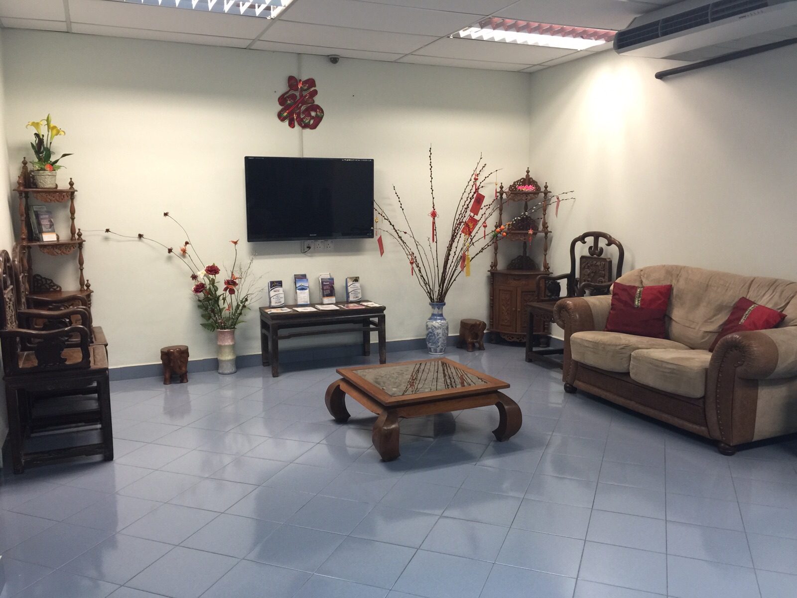
Facilities
Facilities in Chew Eye
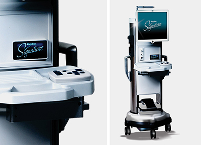
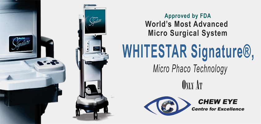
Operating Theatre 1
Cataract
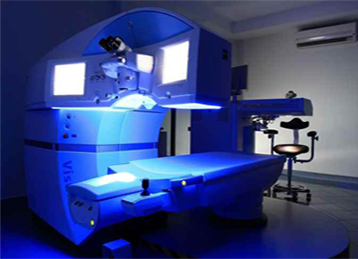
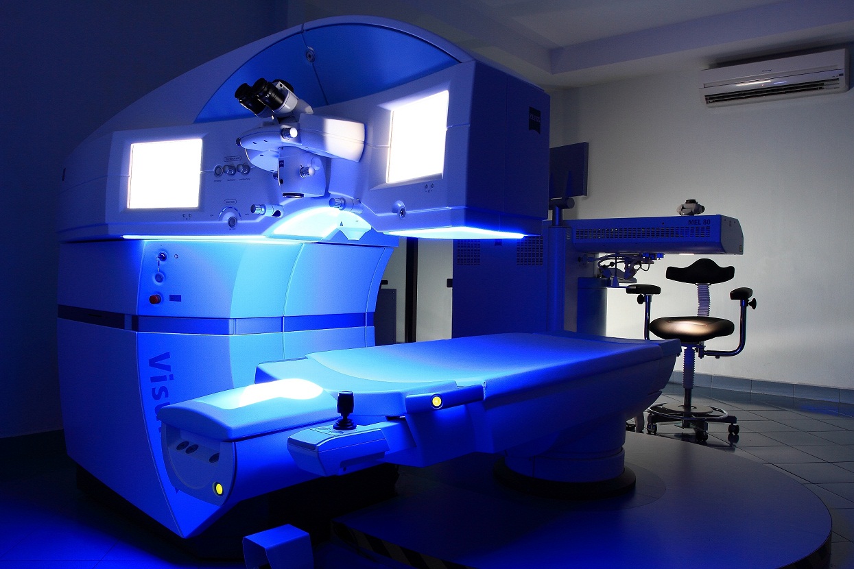
Operating Theatre 2
LASIK
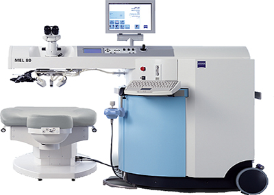
Operating Theatre 3
LASIK & LBV
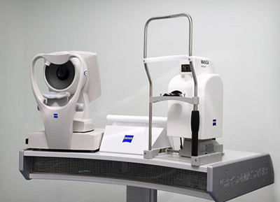
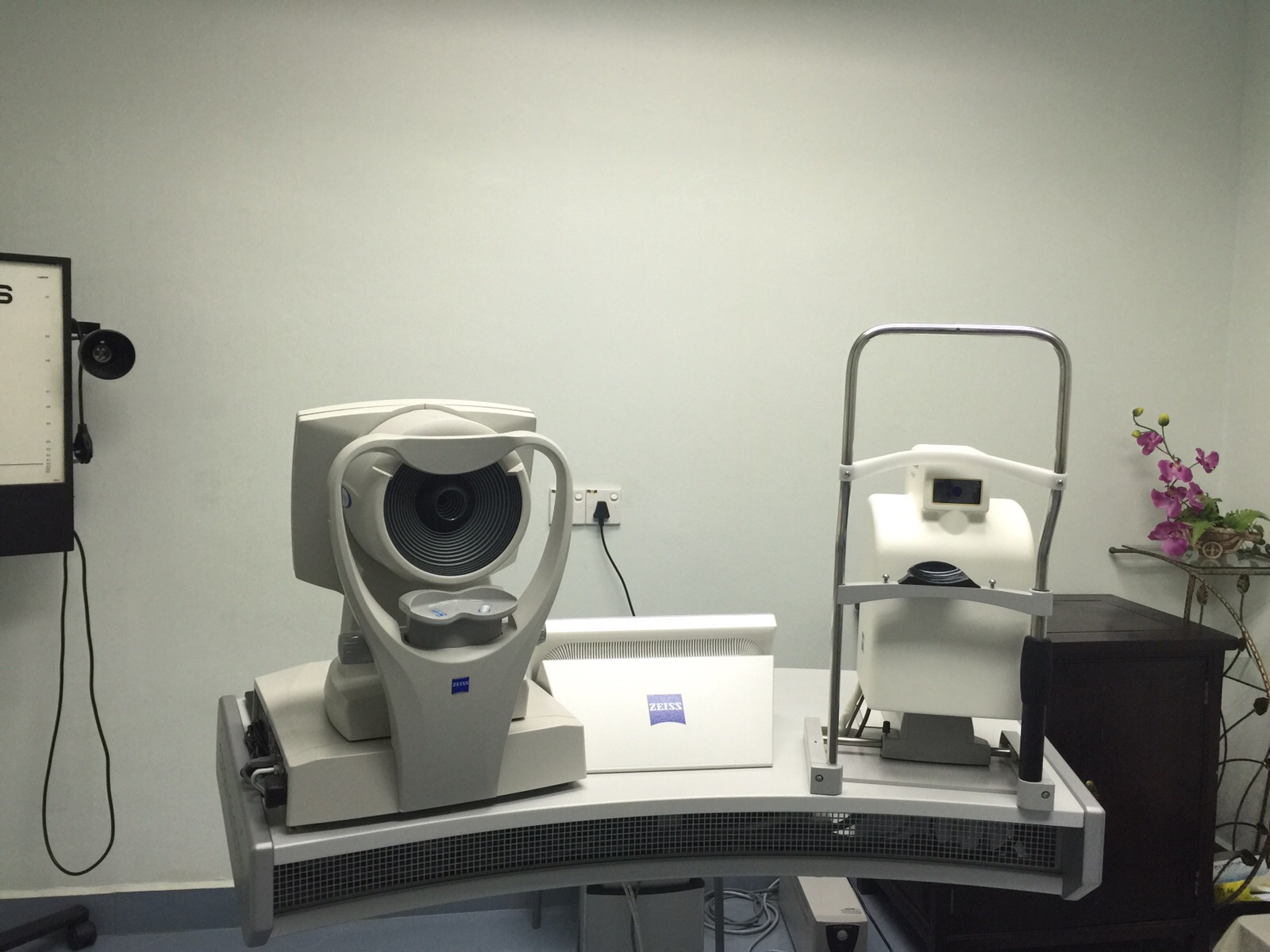
Laser Suite
Corneal Topography & Wasca Analyzer

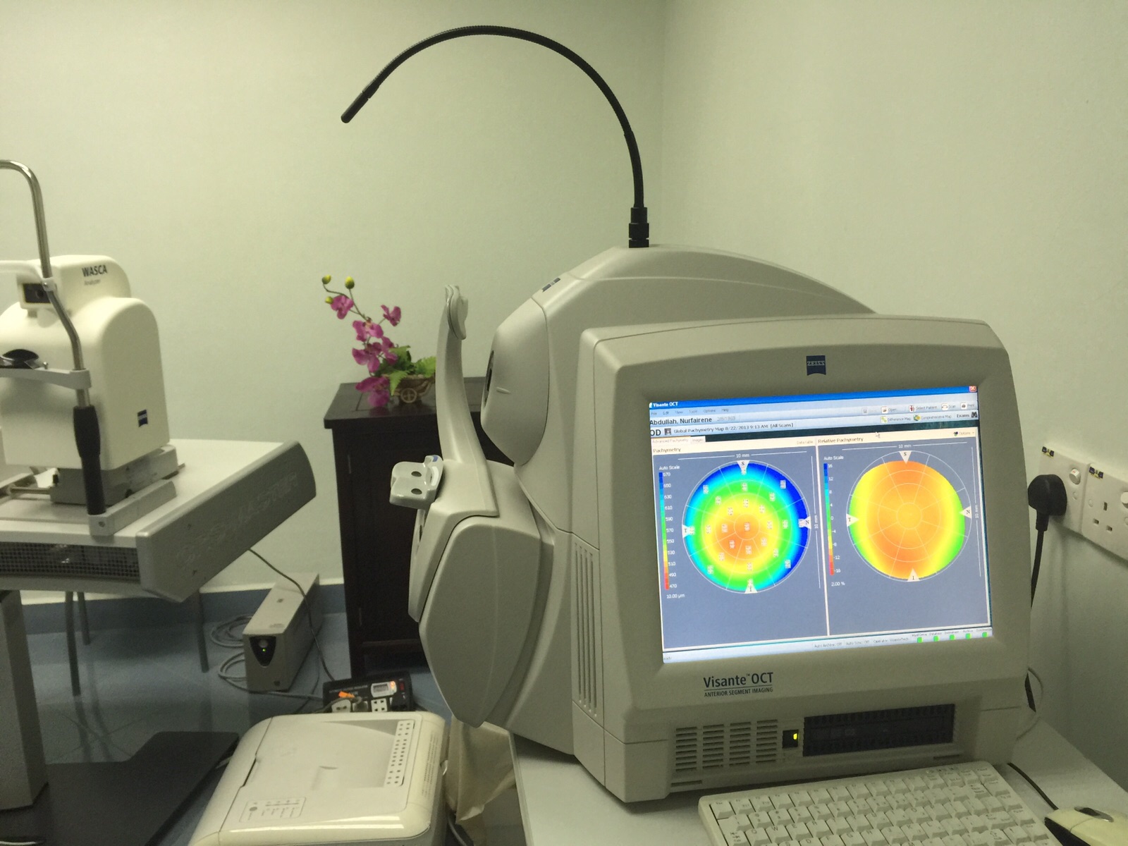
Laser Suite 2
Visante Anterior Segment OCT
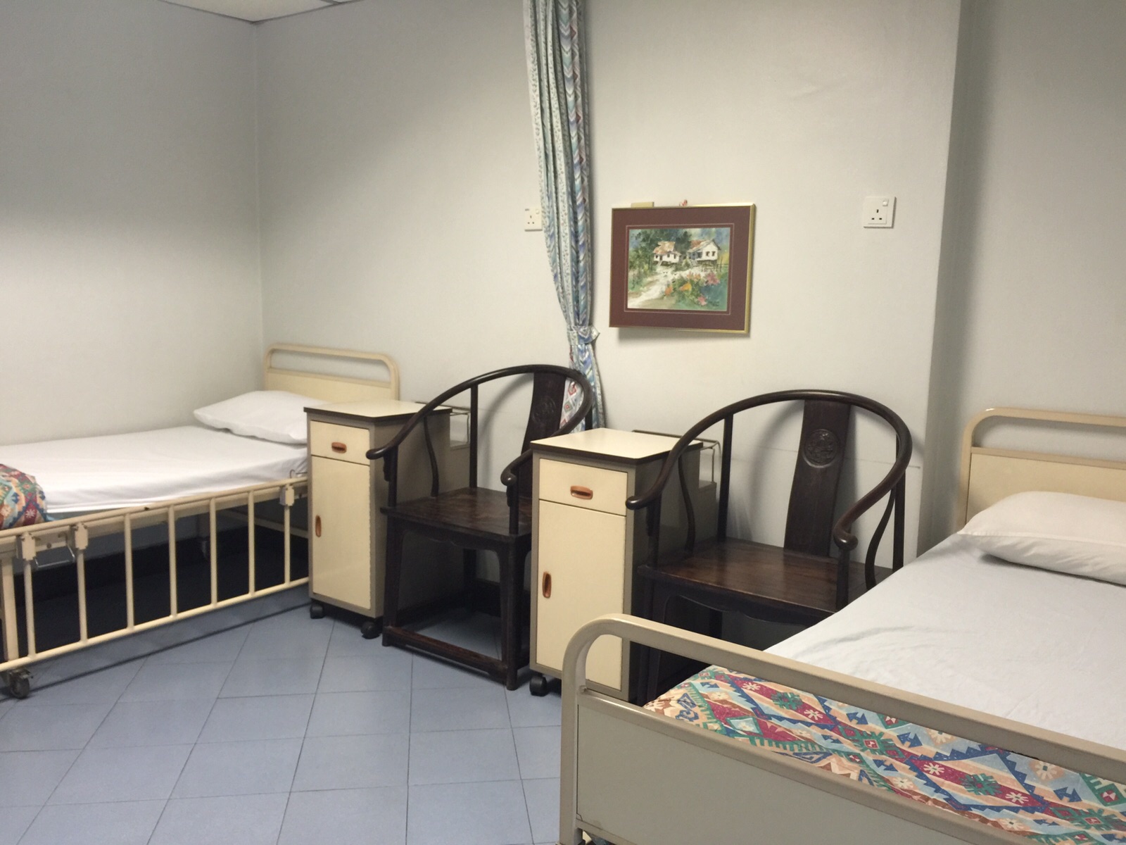
Rooms
Surgery Recovery Rooms

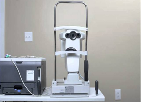
Diagnostic Equipment
IOL Master

Diagnostic Equipment
A-scan Ultrasound Biometry ( E-Z Scan AB5500+)
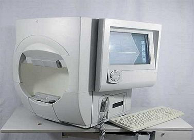
Glaucoma Analysis
Humphrey Field Analyser